How did my cells die? Help choose the right apoptosis assay

How did my cells die? Help choose the right apoptosis assay

Understanding cell death has a huge impact on the treatment of diseases. Apoptosis is a genetically determined suicide program that removes unnecessary or potentially harmful cells. Apoptosis is an important process in embryonic development, cell aging, the immune response and the response to poisoning. Deviations in this program can lead to neurodegenerative or autoimmune diseases. Blocking apoptosis is also one of the “hallmarks of cancer” postulated by Hanahan and Weinberg. 1

Apoptosis leads to a clear morphological change in the cell due to a non-inflammatory cascade of molecular events. These include the reduction of cell volume, the fragmentation of the cell nucleus, the condensation of chromatin, the degradation of DNA, the formation of bubbles on the cell membrane and the subsequent formation of apoptosis bodies. This sequence not only leads to the destruction of unwanted cells, but also prepares the cell debris for removal by phagocytes. In view of the variety of apoptotic stimuli and their effects on different signaling pathways, it is important to understand what exactly led to cell death in an experimental setup.

Mitochondrial membrane potential
Mitochondria are involved in the apoptotic process in several places. During the life of a cell, mitochondria use oxidizable substrates to create a proton gradient along the inner mitochondrial membrane. During apoptosis, this membrane potential decreases in connection with the opening of the mitochondrial permeability pores and the release of apoptogenic factors such as cytochrome C. In some apoptotic models, the loss of membrane potential is considered an early event in the apoptotic process. Other researchers suspect that the loss of membrane potential is a result of the apoptotic signaling cascade. 2-4 An easy way to assess the membrane potential of mitochondria in a cell population is to use positively charged dyes such as JC-1 and TMRE, which are found in the electronegative Collect inside active mitochondria.

JC-1 / TMRE
JC-1 changes from red fluorescence in healthy mitochondria to green fluorescence when, as in apoptosis, the membrane potential is lost. TMRE accumulates in polarized mitochondria and is particularly suitable for observing the membrane potential of living cells. Depolarized mitochondria with reduced membrane potential are unable to accumulate TMRE.

Caspase activation
The key figures in the apoptotic signaling pathway are cysteine-dependent ASpartate-specific ProteASEN (caspases), the efficient activation of which determines the fate of the cell. Caspases are activated by several signal paths and form reinforcing loops. The external signaling pathway binds extracellular death ligands such as FasL or TNF-α to transmembrane death receptors, which in turn recruit initiator caspase-8. In the inner signal pathway, the cytochrome C released by mitochondria can trigger the formation of the caspase-activating complex (also called apoptosome). The latter in turn recruits and activates the initiator caspase-9. The initiator caspases then cleave and activate the effector caspases 3 and 7 and lead to the cleavage of specific substrates, which leads directly to the morphological changes that traditionally define cellular apoptosis.5 This is a measurement of the caspase activity important indicator of the ongoing apoptotic process.

Caspase 3/7
The protease activity of caspases 3 and 7 can be detected using the fluorogenic substrate N-Ac-DEVD-N’-MC-R110, which produces a fluorescent product on cleavage.

Core fragmentation, chromatin condensation and DNA degradation
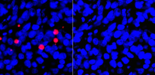
Caspases also break down internal cell structures and help with their efficient disposal. One of the most remarkable events in this process is the condensation of the cell nucleus and its breakdown into smaller fragments. The core fragmentation results from the dissolution of the nuclear lamina after proteolysis by caspases and the collapse of the nuclear envelope. Another characteristic of apoptosis is the condensation of chromatin, accompanied by hydrolysis of the core DNA. Hoechst 33342, a cell-permeable, fluorescent DNA dye, is often used to microscopically analyze chromatin condensation. The Golgi, the endoplasmic reticulum and the mitochondrial network also disintegrate during apoptosis. During the breakdown, the fragments are distributed in bubbles along the plasma membrane.

DNA dyes
Hoechst dyes are cell-compatible and bind nucleic acids in living and fixed cells. Its blue, and therefore little overlapping, emission spectrum makes it recommended for researchers who plan to carry out several fluorescent stains on a sample. An alternative cell-compatible DNA dye is DRAQ5 ™, whose deep red excitation spectrum can be combined with fluorophores that emit blue and orange light. Non-membrane-compatible dyes such as propidium iodide, DAPI, DRAQ7 ™ or RedDot ™ 2 are ideal for the specific staining of the nuclei of dead cells where the integrity of the plasma membrane is impaired.

Blistering
Caspases also cleave many major components of the cytoskeleton, thereby rounding and retracting the cells, which is typical of early stages of apoptosis. Another result of the weakening of the cytoskeleton is the formation of membrane vesicles. When the cytoplasm presses against weakened areas of the plasma membrane, these show themselves as bulges that can be visualized microscopically. These vesicles are believed to be a result of myosin-dependent contraction of cortical actin bundles that press the cytoplasm against the cell cortex. Blistering on the plasma membrane is an important step on the path of the apoptotic cell towards the smaller apoptotic bodies.

Elimination of apoptotic cells
A critical component of the apoptotic process is the complete lack of an inflammatory response to dying cells. The disintegration of the apoptotic cells into apoptotic bodies prevents the release of the Damage-Associated Molecular Patterns (DAMPs) into the extracellular space and thereby facilitates the disposal by phagocytes. In early apoptosis, apoptotic cells secrete a “find me” signal and later a “eat me” signal to recruit phagocytes. This targeted interaction between apoptotic cells and phagocytes ensures that dying cells are eliminated by the non-inflammatory signaling pathway. “Find me” signals that recruit phagocytes include lysophosphatidylcholine, sphingosine-1-phosphate, fractal kinine and nucleotides such as ATP and UTP. These nucleotides are released through a caspase-mediated channel opening of the pannexin-1 (PANX1) channels.6 The caspase-dependent PANX1 channel opening also allows a small group of fluorescent monomeric cyanine dyes such as TO-PRO®-3 to flow in. In this way, the pannexin channels can be used to identify cells that send “find me” signals. 7

As soon as phagocytes approach an apoptotic cell, they recognize phosphatidylserine and phosphatidylethanolamine residues that come from inside the phospholipid double membrane and are now exposed on the cell surface. This rearrangement, which is typical of apoptotic cells, distinguishes it from its viable counterparts and provides the phagocytes with a “eat me” signal. The phospholipid-binding protein Annexin V adheres to phospholipid residues that are exposed during apoptosis. It is used to detect phosphatidylserine on the outer membrane of apoptotic cells and to determine the “eat me” phase of apoptosis.8,9

TO-PRO®-3
TO-PRO®-3 is a deep red fluorescent dye that enables the detection of an early event of apoptosis. The dye uses the caspase-dependent activated pannexin channels to penetrate the cells. This makes it possible to identify apoptotic cells that cannot be detected with the help of traditional markers such as Annexin V due to the process that has just started.

Annexin V
Annexin V can be conjugated with various fluorochromes (e.g., FITC, PE, APC) to stain phosphatidylserine on the outer plasma membrane. The detection with Annexin V can be combined with other markers, such as those for membrane integrity (for example, RedDot ™ 2, propidium iodide, DAPI), in order to distinguish apoptotic from necrotic cells.

Multiplex is the key
The above-mentioned morphological features of the apoptotic signaling pathway can help determine the type of cell death in a model system. However, a single measurement can lead to misinterpretations for certain experimental questions (e.g. the distinction between necrosis and apoptosis). In contrast to single measurements, multiplex assays provide a more complete picture of the apoptotic process. For example, the combined measurement with TO-PRO®-3, Annexin V, TMRE and DAPI allows a simultaneous statement on “Find me” or “Eat me” signals, the mitochondrial membrane potential and the core fragmentation. Multiplex assays thus provide a complex picture of the apoptotic processes and how they affect cells. In addition, markers for early apoptosis (eg TO-PRO®-3) can be used to identify cells that were considered viable due to the limitations of traditional apoptosis markers (eg Annexin V). Muliplex methods thus, by using different markers for the different phases of apoptosis, enable a quantitative classification of the entire cell population into the stages: viable, early apoptosis, late apoptosis, apoptosis bodies and non-cellular debris.

The selection of cell-based assays from Cayman Chemical provide you with flexible and efficient tools for determining the different stages of cell death in your model system. Biomol’s technical support and Cayman’s product developers will be happy to help you choose the cell-based assay that suits you, so that you are able to get the most accurate answers to your research questions.

How Do You Like Your Science, Wet or Dry? How Two Lab Experiences Influence Student Understanding of Science Concepts and Perceptions of Authentic Scientific Practice.

How Do You Like Your Science, Wet or Dry? How Two Lab Experiences Influence Student Understanding of Science Concepts and Perceptions of Authentic Scientific Practice.

This examine examines how two sorts of genuine analysis experiences associated to smoking behavior-genotyping human DNA (moist lab) and utilizing a database to check hypotheses about elements that have an effect on smoking habits (dry lab)-influence college students’ perceptions and understanding of scientific analysis and associated science ideas.

The examine used pre and put up surveys and a spotlight group protocol to match college students who performed the analysis experiences in a single of two sequences: genotyping earlier than database and database earlier than genotyping.

Students rated the genotyping experiment to be extra like actual science than the database experiment, in spite of the truth that they related extra scientific duties with the database expertise than genotyping. Independent of the order of finishing the labs, college students confirmed positive aspects of their understanding of science ideas after completion of the 2 experiences.

There was little change in college students’ attitudes towards science pre to put up, as measured by the Scientific Attitude Inventory II. However, on the idea of their responses throughout focus teams, college students developed extra refined views concerning the practices and nature of science after they’d accomplished each analysis experiences, unbiased of the order through which they skilled them.

How Do You Like Your Science, Wet or Dry? How Two Lab Experiences Influence Student Understanding of Science Concepts and Perceptions of Authentic Scientific Practice.

Non-stop lab week: An actual laboratory expertise for all times sciences postgraduate programs.

At the Portuguese universities, sensible courses of life sciences are normally professor-centered 2-hour courses. This method ends in college students underprepared for an actual work atmosphere in a analysis/scientific laboratory.

To present college students with a real-life laboratory atmosphere, the Non-Stop Lab Week (NSLW) was created within the Molecular Biomedicine grasp program on the University of Aveiro, Portugal.

The distinctive function of the NSLW is its depth: throughout a 1-week interval, college students carry out a subcloning and a protein expression venture in an atmosphere that mimics an actual laboratory. Students work autonomously, and the development of work is dependent upon attaining the each day objectives.

Throughout the three curricular years, most college students thought of the depth of the NSLW an excellent expertise and basic for his or her future. Moreover, after some expertise in an actual laboratory, college students state that each the strategies and the atmosphere created within the NSLW had been just like what they expertise of their present work scenario. The NSLW fulfills a niche in postgraduate college students’ studying, notably in sensible expertise and scientific pondering. Furthermore, the NSLW expertise gives expertise to the scholars which are essential to their future analysis space.

Citizen Social Lab: A digital platform for human behavior experimentation within a citizen science framework.

Citizen Social Lab: A digital platform for human behavior experimentation within a citizen science framework.

Cooperation is likely one of the behavioral traits that outline human beings, nonetheless we’re nonetheless attempting to know why people cooperate. Behavioral experiments have been largely performed to shed mild into the mechanisms behind cooperation-and different behavioral traits.

However, most of those experiments have been performed in laboratories with extremely managed experimental protocols however with limitations by way of topic pool or choices’ context, which limits the reproducibility and the generalization of the outcomes obtained.

In an try to beat these limitations, some experimental approaches have moved human behavior experimentation from laboratories to public areas, the place behaviors happen naturally, and have opened the participation to most people within the citizen science framework.

Given the open nature of those environments, it’s important to determine the suitable knowledge assortment protocols to take care of the identical knowledge high quality that one can acquire within the laboratories. In this text we introduce Citizen Social Lab, a software program platform designed for use within the wild utilizing citizen science practices. The platform permits researchers to gather knowledge in a extra practical context whereas sustaining the scientific rigor, and it’s structured in a modular and scalable method so it can be simply tailored for on-line or brick-and-mortar experimental laboratories.

Following citizen science tips, the platform is designed to inspire a extra basic inhabitants into participation, but in addition to advertise participating and studying of the scientific analysis course of. We additionally overview the primary outcomes of the experiments carried out utilizing the platform to this point, and the set of video games that every experiment consists of.

Finally, we consider some properties of the platform, such because the heterogeneity of the samples of the experiments, the satisfaction degree of contributors, or the technical parameters that display the robustness of the platform and the standard of the information collected.

Citizen Social Lab: A digital platform for human behavior experimentation within a citizen science framework.
Citizen Social Lab: A digital platform for human behavior experimentation within a citizen science framework. Citizen Social Lab: A digital platform for human behavior experimentation within a citizen science framework.

Lab-on-a-Chip: Frontier Science within the Classroom.

Lab-on-a-chip expertise is introduced into the classroom by way of improvement of a lesson sequence with hands-on practicals. Students can uncover the ideas of microfluidics with totally different practicals protecting laminar circulate, micromixing, and droplet technology, in addition to trapping and counting beads.

A fairly inexpensive novel manufacturing approach utilizing scissor-cut and laser-cut lamination sheets is introduced, which supplies good perception into how scientific lab-on-a-chip gadgets are produced. In this fashion highschool college students can now produce lab-on-a-chip gadgets utilizing lamination sheets and their very own lab-on-a-chip design.

We start with a overview of earlier experiences on the usage of lab-on-a-chip expertise in lecture rooms, adopted by an summary of the practicals and initiatives we’ve got developed with scholar security in thoughts. We conclude with an academic state of affairs and a few preliminary promising outcomes for scholar studying outcomes.

Open Up – the Mission Statement of the Control of Impulsive Action (Ctrl-ImpAct) Lab on Open Science.

Open Up – the Mission Statement of the Control of Impulsive Action (Ctrl-ImpAct) Lab on Open Science.

The current paper is the mission assertion of the Control of Impulsive Action (Ctrl-ImpAct) Lab concerning Open Science. As early-profession researchers (ECRs) in the lab, we first state our private motivation to conduct analysis based mostly on the ideas of Open Science.

We then describe how we incorporate 4 particular Open Science practices (i.e., Open Methodology, Open Data, Open Source, and Open Access) into our scientific workflow. In extra element, we clarify how Open Science practices are embedded into the so-known as ‘co-pilot’ system in our lab.

The ‘co-pilot’ researcher is concerned in all duties of the ‘pilot’ researcher, that’s designing a research, double-checking experimental and information evaluation scripts, in addition to writing the manuscript.

The lab has arrange this co-pilot system to extend transparency, scale back potential errors that might happen throughout the total workflow, and to accentuate collaborations between lab members. Finally, we talk about potential options for normal issues that might come up when training Open Science.

Open Up - the Mission Statement of the Control of Impulsive Action (Ctrl-ImpAct) Lab on Open Science.
Open Up – the Mission Statement of the Control of Impulsive Action (Ctrl-ImpAct) Lab on Open Science.

When Citizens Do Science: Stories from Labs, Garages, and Beyond.

This symposium brings collectively, for the first time, a sequence of private narratives about participation in citizen science and an array of commentaries highlighting the novel moral, societal, scientific, philosophical, and coverage implications these narratives reveal.

This symposium consists of twelve private narratives from people who think about themselves to have engaged in citizen science, starting from particular person self-experimentation to the coordination of large, even worldwide, contributions to well being and environmental analysis. The concern additionally consists of three commentaries on these narratives by specialists in human topic analysis, the philosophy of science, and rhetoric and communication in citizen science.

Given the some ways wherein this sort of work challenges our typical classes of regulation, regulation, ethics, and even the conceptualization of who counts as a “scientist” or what counts as “analysis,” this symposium presents concrete examples that we hope will inform and encourage multidisciplinary dialogue. Not solely will these discussions help citizen science in establishing moral buildings, they’ll undoubtedly additionally supply novel views for reconsidering current buildings.

Recent Advances in Transmission Electron Microscopy for Materials Science at the EMAT Lab of the University of Antwerp.

The fast progress in supplies science that permits the design of supplies all the way down to the nanoscale additionally calls for characterization strategies in a position to analyze the supplies all the way down to the identical scale, equivalent to transmission electron microscopy.

As Belgium’s foremost electron microscopy group, amongst the largest in the world, EMAT is constantly contributing to the growth of TEM strategies, equivalent to excessive-decision imaging, diffraction, electron tomography, and spectroscopies, with an emphasis on quantification and reproducibility, in addition to using TEM methodology at the highest stage to unravel actual-world supplies science issues. The lab’s latest contributions are offered right here along with particular case research so as to spotlight the usefulness of TEM to the development of supplies science.

immunoblot

Western Blotting (also referred to as immunoblotting) is a technique used for analysis of individual proteins within a protein mix

(e.g. a cell lysate). In Western blotting (immunoblotting) the protein mix is applied to a gel electrophoresis at a carrier matrix (SDS-PAGE, native PAGE, isoelectric focusing, 2D gel electrophoresis, etc.) to sort the proteins by dimension , charge, or other differences in individual protein bands. The separated protein bands are then transferred to a carrier membrane (e.g.

nitrocellulose, nylon or PVDF). This process is known as blotting. As they’ve been separated due to interactions with fees, the proteins stick to the membrane in the exact same routine. The proteins within this immunoblot are then available for antibody binding for detection.

The antibodies are conjugated with fluorescent or radioactive tags or enzymes that give a subsequent response with an applied reagent, leading to a coloring or emission of light, enabling detection.


The term Western Blotting is based on a play of words. The southern blot, that can be a method to detect DNA sequences, is named after Ed Southern, who first described this process.

The western blot (immunoblot), in addition to the northern blot (for RNA detection), play the significance of this name.

GPR160 Lentiviral Vector Human pLenti GIII

Product Records
Insert Size 4206 bp

Are designing primers using a Kozak sequence demanded or is ATG enough?


Why is my lentiviral infection of lower efficacy than transduction?
Product Application

How should I store my lentivirus?


Guarantee abm guarantees that the proper ORF construct is provided along with the mRNA expression is displayed upon successful transduction. If that is not the case, we will offer a one-time replacement. Clients must supply adequate data to reveal 80% transfection efficiency using a positive control, and additional qPCR information to evaluate the level of gene expression. The replacement won’t be covered by exactly the identical guarantee.


Stability 1 year when stored at -20°C or reduced in a non-frost freezer.
What beginning variety of puromycin should I use for the antibiotic selection?
Package into Lentiviral particles for top efficiency transduction and stably integrated saying
How are your DNA pLenti vectors provided?
No References Found for Product

I also need an empty vectors for a control, how do I go about ordering this?
What’s the cell density which the client should use for lentivirus infection?
Disclaimer for transcript variants: The supplied accession number refers to the transcript (mRNA) arrangement for this particular product. This clone’s molecular sequence aligns as a point of reference. But, individual transcript sequences of the identical receptor can differ through naturally occurring variations (e.g. polymorphisms), each with its own existence that is valid. This clone is in agreement with the reference, but a review of all variants that are prevailing is recommended. All sales are final.

Other

Choice Marker Bacterial: Kanamycin. Mammalian: Puromycin
What is the difference between the pLenti-III and the pLenti-GIII vectors?
FAQs
The UTX expression will be driven by a CMV promoter, with a GFP reporter. Inserts are flanked by and can be excised using NheI and BamHI provided that inserts do not include any inner NheI or BamHI websites.
Which lentiviral genes have been eliminated from the viral vector to leave it replication deficient?

  1. Disclaimer for extra nucleotides: Cloning can lead to the insertion of nucleotides at the 5′ or 3′ end of the gene of interest which, in most cases, is benign to the stability/functionality and the expression of the gene.

Species Human

Direct non-viral plasmid transfection for immediate expression
Important Considerations for Lentiviruses
What is the quantity provided?
Could you please advice on a protocol for vector extraction out of filter paper?
Enriched Lentivirus Safety Features: Replication Incompetency
Product References

Lentivector Packaging Protocol

Viral Packaging abm recommends utilizing Cat# G074, Lentifectin™ Together with one of the following packaging combinations. The packaging mix for this particular vector is # LV003. Alternatively, you can utilize # LV053 to Cat
Where is the polyA signal in your vectors?
Are they high copy or low copy plasmids? What’s anticipated DNA return?
Does your lentiviral strategy comprise the WPRE gene? In that case, is this the wildtype WPRE sequence?

  1. Disclaimer for planned usage: All of abm’s vectors and viral particles are for research use ONLY and NOT for therapeutic/diagnostic software. abm isn’t responsible for any repercussions arising from the use of its vector(s) from therapeutic/diagnostic program (s).
    Please note that because of the large number of factors related, any further expression analysis (e.g. protein saying ) isn’t covered by the guarantee, as such analysis depends upon the end consumer’s experimental problems.
    Viral Vector Brochure
    Selection-Drug Killing Curve

Format Lentiviral Plasmid

Supporting Protocol
Suggested MOI for Common Cancer Cell Lines
How do I compute the MOI?
Procedure Overview

  1. Disclaimer for gene arrangement: The supplied accession number refers to the transcript (mRNA) arrangement for this particular item. Please verify that this is the desired transcript order by cross-referencing. This is crucial because one gene can have multiple transcripts owing to naturally occurring variations. All sales are final.

Which packing system should I use, 2nd or 3rd generation?
What is the HA tag sequence?

  1. Disclaimer for the stop codon: The stop codon for the inserted gene is situated after the 3′ cloning site in our GIII vectors. Our GIII vectors contain open ORF inserts (no stop codon) to allow for subsequent insertion into additional vectors with C-terminal tags or C-terminal fusion proteins if desired. Please be aware if no mix or label protein is necessary in vectors that a stop codon will need to be inserted by PCR towards the end of the gene insert.
    Do your pLenti vectors incorporate a chimeric RSV promoter upstream of the 5′ LTR?

Vector Size 8829bp

Can you have viral particles for this item?
Plasmid Amplification Protocol

For the GIII third generation lentiviral vectors, are there recombination sites flanking the insert sequence?

Vector pLenti-GIII-CMV-GFP-2A-Puro

BlazeTaq One-Step

BlazeTaq One-Step SYBR Green RT-qPCR Kit (100 qPCR Responses )

A. Amplification curves of GAPDH and Actin respectively in HeLa cells by RNA infusion ranging from 100 ng to 0.1 pg; B. Amplification curves of GAPDH and Actin respectively in HeLa cells by RNA infusion ranging from 100 ng to 0.1 pg; C. Melting curves of the amplified fragments of GAPDH (reddish ) and Actin (blue).


Identification of the level of Actin and GAPDH extract from HeLa cells.


• One-step reactions to measure the relative Quantity of RNA
User Guide
CatNumber: QP070-GC

Comparison of the functionality of BlazeTaq™ RT-qPCR kit with rival ´s merchandise.
BlazeTaq™ One-Step SYBR® Green RT-qPCR kit employs reverse transcriptase to convert RNA into cDNA, and also an antibody-modified Taq DNA polymerase to steer clear of polymerase action before thermal cycling. The antibody dissociates and activity of the Taq polymerase is revived upon heating for 3 minutes. In addition, the buffer system enables amplification efficiency and specificity, in addition to enhancing sensitivity of real-time PCR reactions .


• High amplification performance over broad GC-content Variety
Picture
• High specificity, together with a minimal degree of primer-dimer and non invasive product formation


A. Amplification curves of GAPDH in HeLa cells by RNA infusion ranging from 100 ng to 0.1 pg using BlazeTaq™ (reddish ) and Competitor P´s kit (blue); B. Melting curves of amplified fragments of GAPDH with BlazeTaq™ (reddish ) along with Competitor P kit (blue). Amplification is shown by the summit from rival.

Melt peak


Comparison of the operation of BlazeTaq™ RT-qPCR kit with rival ´s merchandise.
Performance
Picture
Cost: 187 $
A. Amplification curves of GAPDH in HeLa cells by RNA infusion ranging from 100 ng to 0.1 pg with BlazeTaq™ (reddish ) and Competitor T´s kit (blue); B. Amplification curves of Actin from HeLa cells by RNA infusion ranging from 100 ng to 0.1 pg using BlazeTaq™ (reddish ) along with Competitor T´s kit (blue).
Provider: GeneCopoeia

qRT-PCR SuperMix Kit

RT-qPCR Kits for RT-qPCR, or quantitative reverse transcriptase PCR or Real Time RT-PCR, Empower quantitative PCR Straight from RNA samples.

These ready-to-use kits include the necessary components for carrying both cDNA synthesis and qPCR. Kit components typically consist of reverse transcriptase, polymerase, buffer, fluorescent probes, and dNTPs. Added reagents such as RNase inhibitors and enhancers may also be contained.

cDNA

For a streamlined procedure that combines cDNA synthesis and qPCR in one reaction tube, consider a”one-step” structure.

Gene-specific RT-qPCR assay kits are also available, which include prepackaged primer places. Visit the provider page for details.

cDNA-Random Hexamer

cDNA-Random Hexamer

One-step RT-PCR reactions consistently use a gene-specific primer for first-strand cDNA synthesis, while two-step RT-PCR reactions permit for additional priming choices.


“When the mRNA includes a poly-A 3′ tail, then a oligo-dT primer may be used to prime each of mRNAs simultaneously.
SSPs give the best specificity and also have been proven to be the most persistent of the primer selections for reverse transcription. But they don’t offer you the flexibility of oligo(dT) and random primers, meaning a fresh cDNA synthesis reaction has to be done for each gene to be analyzed. This creates primers for processing restricting cell or tissue samples.
Oligo(dT) primers aren’t recommended as the sole real primer for cDNA synthesis of 18S rRNA can be used for normalization at a real-time PCR experimentation as the oligo(dt) primer won’t anneal.


To create cDNA in the subset of mRNA, afterward a sequence-specific primer can be utilized that will only bind to a mRNA sequence.

Multiple kinds of oligo(dT) primers can be found. Finally, anchored oligo(dT) primers are made to steer clear of polyA slippage by making sure they anneal in the three ´UTR/polyA junction. Deciding on the most effective oligo(dT) primer can rely in part on the warmth of the reverse transcription. Thermostable RTs like SuperScript® III Reverse Transcriptase may function better with primers, which stay annealed in comparison for their counterparts.
Sequence Particular Primers
http://www.bio.davidson.edu/courses/genomics/method/cdnaproduction.html
Implementing a mix of random and oligo(dT) primers can at times raise information quality by combining the advantages of both if utilized in precisely the exact same first-strand cDNA synthesis reaction. Random primers are utilized in reactions.


Oligo(dT) primers are a favourite choice for two-step cDNA synthesis reactions due to their specificity for both mRNA and since they permit several distinct targets to be analyzed by precisely the exact same cDNA pool. Since they initiate transcription structure that is hard can lead to cDNA synthesis.

In case you wanted to create parts of cDNA which were scattered all around the mRNA, then you may use an arbitrary primer cocktail which could create cDNA from many mRNAs however, the cDNAs wouldn’t be full length. The advantages to priming would be the creation of cDNA fragments that are shorter and raising the likelihood that 5′ ends of the mRNA will be converted into cDNA. Since reverse transcriptase does not usually achieve the 5′ ends of extended mRNAs, arbitrary primers may be beneficial”

Since they anneal through the target molecule They’re also excellent for RNA, such as RNA.